Recent studies of the literature present models of the tumor microenvironment to study how physics affects tumors and revealed that cell movements are governed by mechanical forces of interaction between cells and between cells and the extracellular matrix. The adhesion capacity of cancer cells to the stroma that surrounds them induces intracellular contraction forces that deform their microenvironment through the alignment of collagen fibers, altering its mechanical properties. Thus, we intend in this Project to study the effects of intercellular and cell-ECM interactions through a new technological strategy based on the use of low intensity ultrasound combining bioprinted models, 2D and 3D cell samples, as well as ex-vivo and in vivo samples of mouse. The key advantage of 3D printing cancer cells is the potential to model the tumor microenvironment in-vitro with very high fidelity, offering a greater representation of tumor formation and progress to analyze its response to drugs and avoiding the use of animal samples
building up!!
Selection and Optimization of a Bioink Based on PANC-1-
Plasma/Alginate/Methylcellulose for Pancreatic TumourModelling
Cristina Banda Sánchez, Nieves Cubo Mateo, Laura Saldaña, Alba Valdivieso, Julie Earl,
Itziar González Gómez and Luis M. Rodríguez-Lorenzo
Polymers 2023, 15(15), 3196; https://doi.org/10.3390/polym15153196
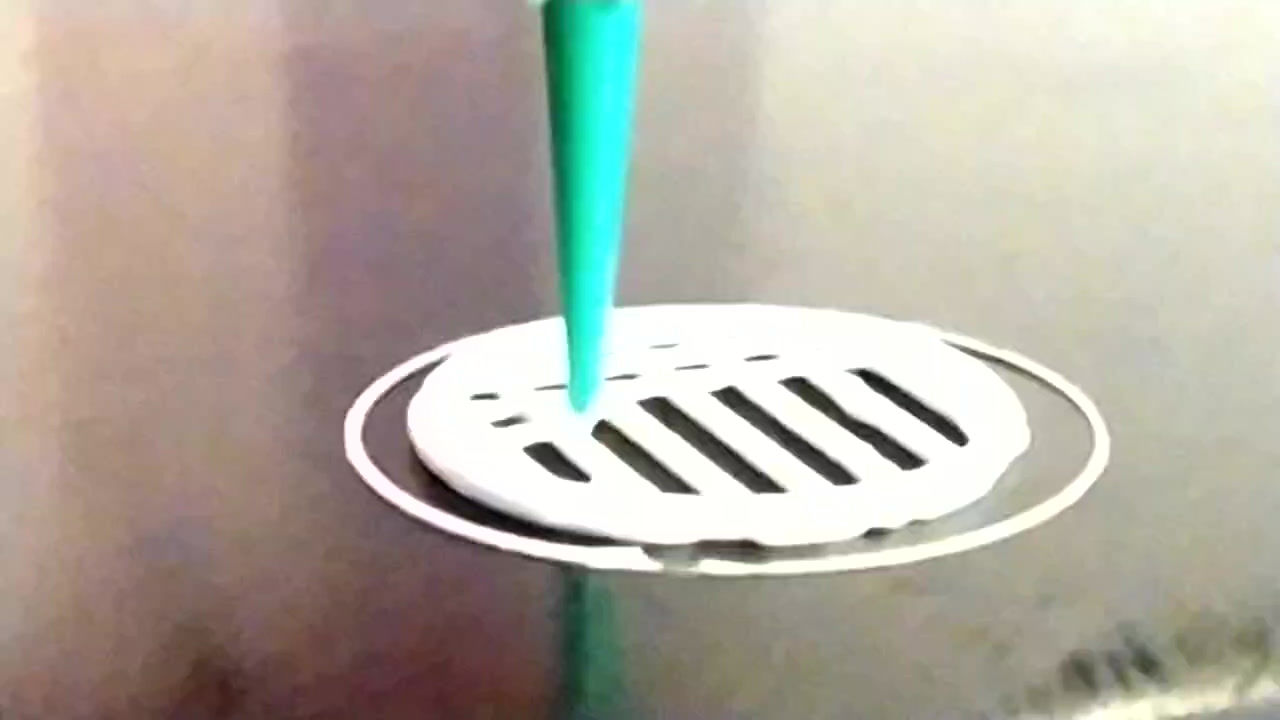
3D bioprinting involves using bioinks that combine biological and synthetic materials. The
selection of the most appropriate cell-material combination for a specific application is complex,
and there is a lack of consensus on the optimal conditions required. Plasma-loaded alginate and
alginate/methylcellulose (Alg/MC) inks were chosen to study their viscoelastic behaviour, degree of
recovery, gelation kinetics, and cell survival after printing. Selected inks showed a shear thinning
behavior from shear rates as low as 0.2 s1, and the ink composed of 3% w/v SA and 9% w/v MC
was the only one showing a successful stacking and 96% recovery capacity. A 0.5 106 PANC-1
cell-laden bioink was extruded with an Inkredible 3D printer (Cellink) through a D = 410 m tip
conical nozzle into 6-well culture plates. Cylindrical constructs were printed and crosslinked with
CaCl2. Bioinks suffered a 1.845 Pa maximum pressure at the tip that was not deleterious for cellular
viability. Cell aggregates can be appreciated for the cut total length observed in confocal microscopy,
indicating a good proliferation rate at different heights of the construct, and suggesting the viability
of the selected bioink PANC-1/P-Alg3/MC9 for building up three-dimensional bioprinted pancreatic
tumor constructs.
